|
NOAA Gordon Gunter last revised 25-May-2002
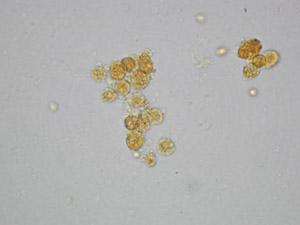
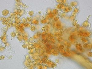
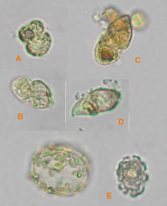
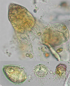
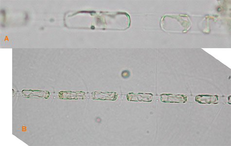
THE UNKNOWN GREEN FROM THE DEEP To determine the origin of a shallow subsurface chlorophyll maximum at ca. 50 m depth encountered at some stations along the cruise track in the northern/central part of the survey area, two samples were provided: a net sample from the Mocness of stn. 104 (5/6/2002) from the depth horizon 25-50 m; and a water bottle sample from stn. 95 (5/5/2002) from 58 m depth (chlorophyll maximum). For the location of these stations, refer to the cruise protocols. It was my understanding that both stations were outside the center of the immense shallow subsurface maximum. Both samples were preserved by Lugol's Iodine and studied by light microscopy. All photographs taken at 400x magnification on a Zeiss Axioskop equipped with a Nikon CoolPix 990 digital camera. The Mocness sample contained numerous clumps of spherical cells, which appeared to be in the process of degradation, in part highly covered by bacteria, and of dinoflagellate origin (Figs. 1, 2). The cell clumps were entangled in a transparent organic matrix and zooplankton, predominantly chaetognaths. Despite their unhealthy look, cells still contained chlorophyll that may have produced the subsurface chlorophyll maximum signal of the in-situ CTD probe. The water bottle sample (phytoplankton was enriched in Utermöhl sedimentation chambers for inspection) contained the same spherical cells. In addition, Gymnodinium-like dinoflagellates were quite abundant (Fig. 3A-C), as were oligotrich ciliates (Fig. 4). Amphidinium (Fig. 3D), coccolithophorids (Fig. 3E), and the diatoms Skeletonema sp. (Fig. 5A) and Chaetoceros sp. (Fig. 5B) were found in small numbers. Small forms of the diatom Rhizosolenia, which were discussed in relation to the Florida black waters, occurred only sporadicly.
|