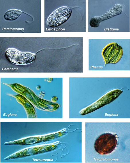
- Characteristic genus: Euglena
- Abundant in almost any nutrient-rich freshwater system
- Dissolved organic material fosters euglenophyte abundance
- Require vitamins B1 and B12 auxotrophic species
- Pigments: Chl.a, Chl.b, b-carotene, diadinoxanthin
- Secondary plastids; some species possess colorless plastids or lost their plastids; these species are phagotrophic
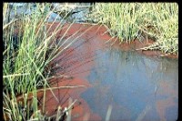
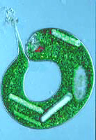
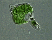
- Color of most species green, but some produce red water blooms
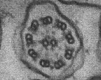
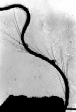
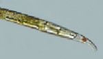
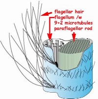
- One or two (visible) flagella of typical eukaryotic 9+2 structure
- Evolutionary probably the oldest group of eukaryotic algae
- Unicellular motile or sessile forms
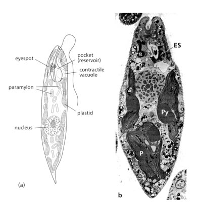
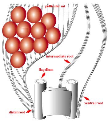
- Flagella: 2 flagella emerge from a pocket at the cell anterior (reservoir, ampulla); sometimes only 1 flagellum extends outside the pocket; flagella carry one row of long hairs and shorter hairs on flagella surface
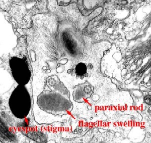
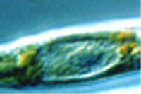
- Phototrophic species possess an eye-spot made of carotenoids (directed swimming to light source = phototaxis)
- Secondary plastids show 3 membrane layers
- Storage product: paramylon, does not stain blue-black with iodine
- Euglenophyte Flagellar Apparatus
- Ampulla evolved from separate flagella and the cytostome (cell mouth)
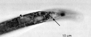
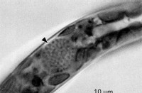
- Contractile vacuole: adjacent to ampulla; discharges excess water into ampulla
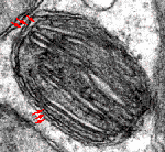
- Paraflagellar rod: protein structure at the roots of flagella, makes flagella appear thicker, involved in flagella motion control
- Flagellar roots: bands of microtubules from flagella bases to cytoplasm, act like muscles (control cell shape); striated connective between flagella bases coordinates flagellar motion

Euglenophyte Swimming
- Swim by one or two flagella; only the tip of flagellum is moving, propelling the cell forward
- Light sensing system: 2 major parts,
- paraflagellar rod at the base of at least the emergant flagellum; contains light-sensitive flavins
- Eye-spot (stigma): in the cytoplasm adjacent to the ampulla; bright orange by carotenoids
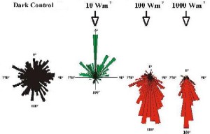
- Light direction: the eye-spot shades the paraflagellar body while cell is swimming in rotating movements along its axis
- Positive phototaxis: most photo- trophic euglenophytes swimm towards the light source
see the heterotrophic euglenophyte Peranema swim (QuickTime 960 kB)
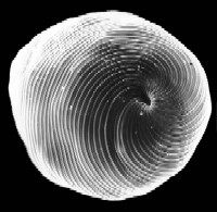
- Cell wall is called pellicle, made 70-80% protein plus lipids
-
Pellicle
is organized in strips, long ribbons that extend helically along the cell;
the edges of the ribbons are bent upwards and downwards, resp.
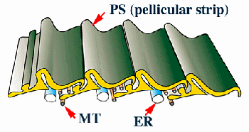
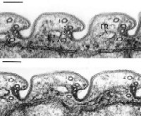
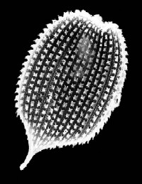
Upper: schematic drawing of the euglenophyte pellicle, showing four strips with their bent edges and rows of four microtubuli; middle: cross section (EM) through the pellicle (left) and a Euglena cell (right); lower: scanning electron micrographs of the pellicle of Euglena (left) and Phacus (right).
- Microtubuli at the edge of the stripes faciliate lateral sliding of strips past each other
-
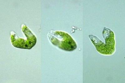
Metaboly:
flexible movement of the pellicle cause change of cell shape; typical for
euglenophytes only; allows to stem through sand grains, etc.
 See also
mataboly QuckTime (1.3 MB)
See also
mataboly QuckTime (1.3 MB) - Cell division: prior to cell division, pellicle strips are doubled
- Mucilage: euglenophytes can excrete polysaccharids or glycoproteins from mucocysts. Mobile cells have only thin mucilage layer, but immobile, round cells can aggregate in thick jelly layers: palmella or palmelloid stages
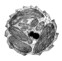
Euglenophyte cysts (left); stained mucocysts in Euglena (right)
Reproduction
-
Asexual
reproduction by longitudinal cell division
starting from the front end of the cell
-
DNA
is permanently condensed, i.e. no cell cycle changes in coiling
- Mitosis: nucleus moves towards the ampulla/reservoir; mitosis occurs within nucleus envelope
- Sexual reproduction: unknown
-
Cysts
are formed to survive unfavorable conditions; thick, mucilaginous cell
wall, loss of flagella, eye-spot mostly present, rounding of cells, increase
in paramylon granules
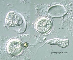
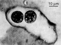
Germinating cysts of the heterotrophic, colorless Pernanema